Grant number: 2019/33/B/NZ7/02980
Principal Investigator: Małgorzata Brindell
Source of funding: National Science Center Poland
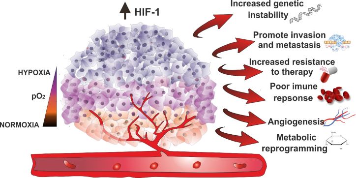
A common feature of most tumors is lowered levels of oxygen. Such a non-physiological level of oxygen is called hypoxia. This state is a consequence of an imbalance between the supply and consumption of oxygen. In addition, hypoxia could promote tumor invasion and metastasis as well as the resistance of tumor cells to therapy. Progressive hypoxia is prognostic for poor patient outcomes.
 There is an urgent need for the development of sensors, that have an increased response to subtle variations in oxygen concentration, increased kinetic stability, and accumulate only in the hypoxic cancer tissues, avoiding the necrotic or normoxic ones and have a detection system that is relatively cheap, portable and easy to operate. To fulfill all these requirements, we have designed a group of sensors based on the conjugation of bioreductive compounds with fluorescent units to form a new type of hypoxia-selective optical imaging sensor.
This project aims to select the compounds that allow for the optical identification/detection of the hypoxic tissue, including oxygen evaluation within it. As an outcome, the collected information should allow for a rational selection of compound/s which might be further tested for its/their clinical application and can be used as a probe in molecular biology studies.
E. Janczy-Cempa, O. Mazuryk, D. Sirbu, N. Chopin, M. Żarnik, M. Zastawna, C. Colas, M.-A. Hiebel, F. Suzenet, M. Brindell
Sensors & Actuators B Chem. 2021, 346, 130504
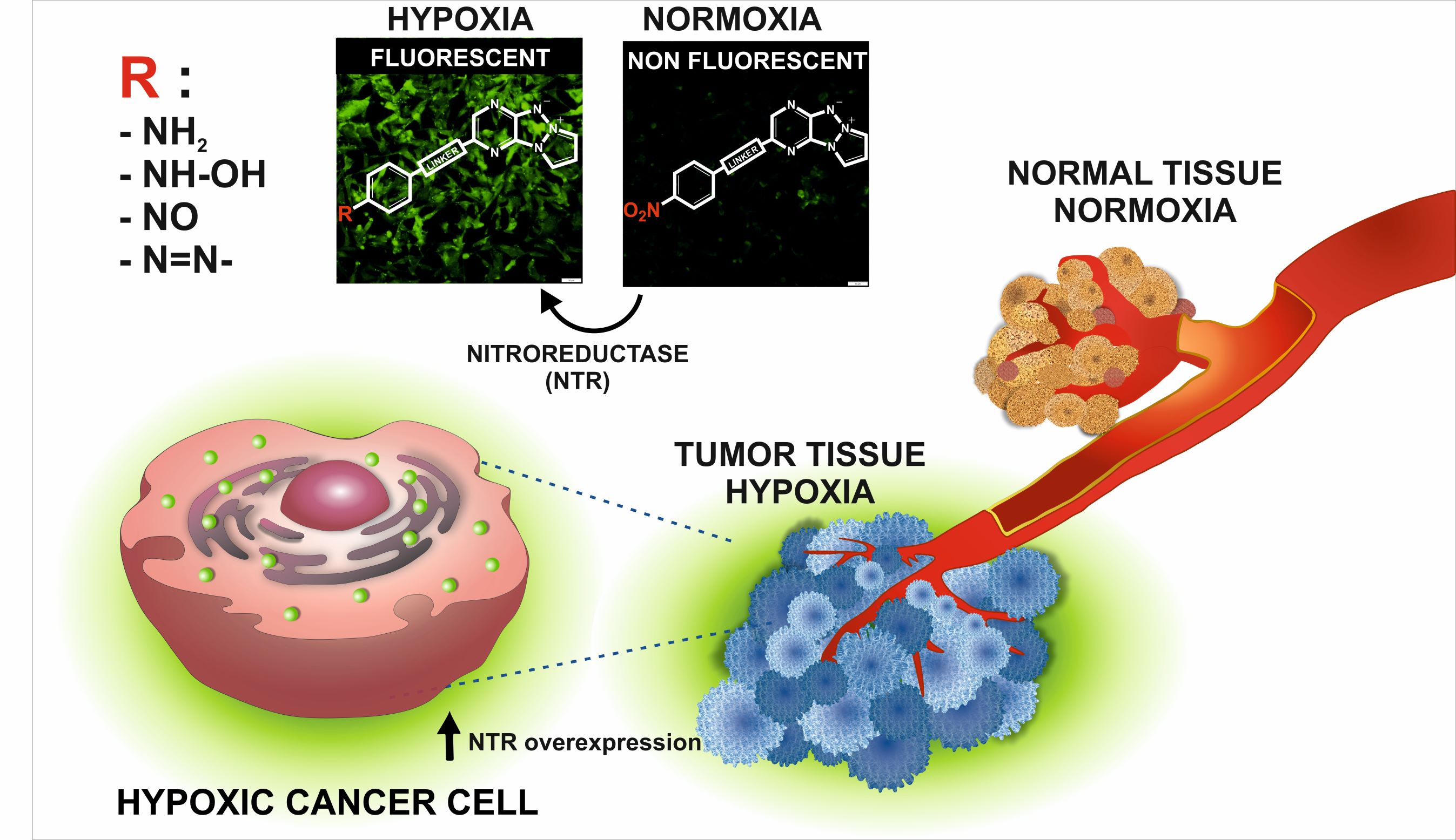
Nitroreductases (NTRs), a family of flavin-containing enzymes, can be overexpressed in regions of tumor hypoxia, that is, areas deprived of oxygen. The detection of NTRs may be applied to monitor the hypoxia level in tumors. To quantify NTRs, novel sensors were designed based on the conjugation of pyridazino-1,3a,6a-triazapentalene to a para-nitrophenyl. They had a weak fluorescence (form off) whereas their reduction by NTRs led to the 15-fold enhancement of fluorescence intensity (form on). The reaction was sensitive and selective and the formation of products was directly proportional to the amount of the enzyme. The in vitro studies showed that probes readily accumulated in cells without exhibiting host cell toxicity. The designed compounds allowed for fast determination of the difference in NTR content of cells without the need for special sample preparation such as cell lysis. Changes can be monitored using a plate reader, flow cytometer, and fluorescence microscope making these probes a remarkably universal tool.
E. Janczy-Cempa, O. Mazuryk, A. Kania, M. Brindell
Cancers 2022, 14, 12686
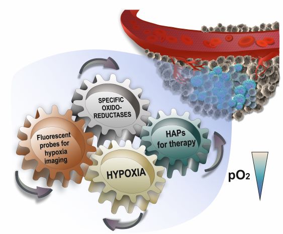
The review focuses on current knowledge about the expression and activity of oxidoreductases which are relevant in the activation of hypoxia-activated prodrugs (HAPs) and fluorescent imaging probes. The current clinical status of HAPs, their limitations, and ways to improve their efficacy are briefly discussed. The fluorescence probes triggered by reduction with specific oxidoreductase are briefly presented, with particular emphasis on those for which correlation between the signal and enzyme expression determined with biochemical methods is achievable.
E. Janczy-Cempa, A. Kwiatkowska, O. Mazuryk, N. Chopin, M.-A. Hiebel, F. Suzenet, M. Brindell
Sensors & Actuators B Chem. 2023, 394, 134450
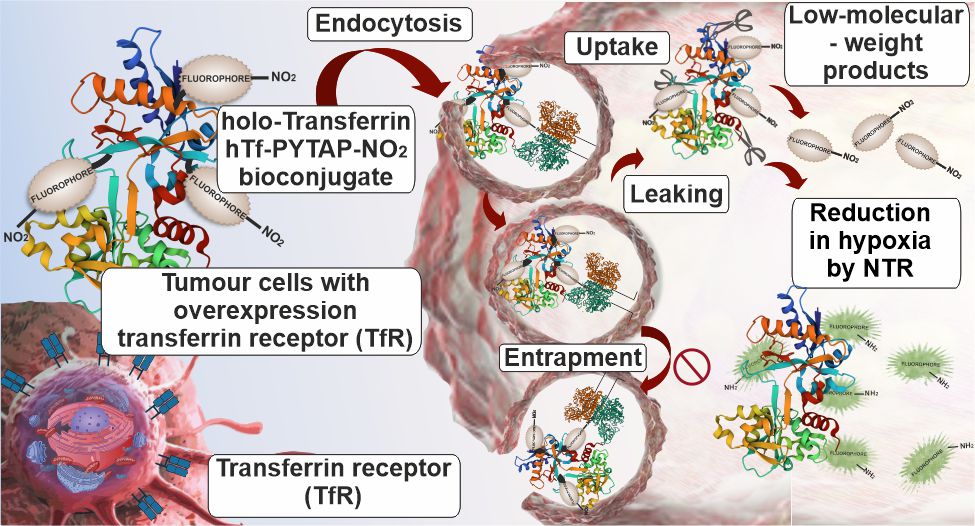
A new fluorescent sensor for imaging cancer cells was designed by bioconjugation of holo-transferrin (hTf) with a nitro-pyrazinotriazapentalene moiety (PyTAP-NO2) as a bioreductive fluorescent unit. hTf was chosen to increase its selective delivery to cancer cells through transferrin receptor (TfR) mediated endocytosis. PyTAP-NO2 was applied to enhance the fluorescent signal in oxygen-deprived cells due to the increased expression of nitroreductases under such conditions. A protein-dye bioconjugate had an average of 8 fluorophore molecules per protein that effectively amplified the fluorescence signal. The designed hTf- PyTAP-NO2 was non-toxic and its efficient accumulation through TfR, as well as its high stability and sustained intracellular retention, was proven. hTf-PyTAP-NO2 bioconjugate allows selective accumulation in cancer cells containing increased levels of TfRs with the simultaneous slight enhancement of signal in hypoxic cells due to increased expression of nitroreductases.